6.5.1 Genetic Engineering – Video
6.5.1 Genetic Engineering
By: ZogScience
See original here:
6.5.1 Genetic Engineering - Video
Recommendation and review posted by Bethany Smith
BIOLOGY: Genetic Engineering – Robin Hesketh – Video
BIOLOGY: Genetic Engineering - Robin Hesketh
Good News for cancer therapy. Trials show genetic engineering of T-cells in the treatment of B-Cell Acute Lymphocitic Leukemia works.
By: Hay Levels
See the rest here:
BIOLOGY: Genetic Engineering - Robin Hesketh - Video
Recommendation and review posted by Bethany Smith
Future Gene Therapy for Cancer – Video
Future Gene Therapy for Cancer
Future Gene Therapy for Cancer. Video streamed by http://www.AllthingsScience.com.
By: Davies Robinson
Originally posted here:
Future Gene Therapy for Cancer - Video
Recommendation and review posted by Bethany Smith
How mutant gene can cause deafness
Scientists at The Scripps Research Institute (TSRI) have discovered how one gene is essential to hearing, uncovering a cause of deafness and suggesting new avenues for therapies.
The new study, published November 20 in the journal Neuron, shows how mutations in a gene called Tmie can cause deafness from birth. Underlining the critical nature of their findings, researchers were able to reintroduce the gene in mice and restore the process underpinning hearing.
"This raises hopes that we could, in principle, use gene-therapy approaches to restore function in hair cells and thus develop new treatment options for hearing loss," said Professor Ulrich Mller, senior author of the new study, chair of the Department of Molecular and Cellular Neuroscience and director of the Dorris Neuroscience Center at TSRI.
The Gene Responsible
The ear is a complex machine that converts mechanical sound waves into electric signals for the brain to process. When a sound wave enters the ear, the uneven ends (stereocilia) of the inner ear's hair cells are pushed back, like blades of grass bent by a heavy wind. The movement causes tension in the strings of proteins (tip links) connecting the stereocilia, which sends a signal to the brain through ion channels that run through the tips of the hair cell bundles.
This process of converting mechanical force into electrical activity, called mechanotransduction, still poses many mysteries. In this case, researchers were in the dark about how signals were passed along the tip links to the ion channels, which shape electrical signals.
To track down this unknown component, researchers in the new study built a library of thousands of genes with the potential to affect mechanotransduction.
The team spent six months screening the genes to see if the proteins the genes produced interacted with tip link proteins. Eventually, the team found a gene, Tmie, whose protein, TMIE, interacts with tip link proteins and connects the tip links to a piece of machinery near the ion channel.
A Path to New Treatments
This discovery answers a long-standing question in neuroscience. Scientists have long known that mutations in the Tmie gene could cause deafness -- but they weren't sure how.
See the original post:
How mutant gene can cause deafness
Recommendation and review posted by Bethany Smith
Doctors working on gene therapy to help patients with hemophilia
SOUTH BEND, Ind.--- Little Hunter Miller's motor is always running.
Like most toddlers he's sometimes one step away from trouble, but for Hunter being rough and tumble can have serious side effects. Hunter has severe hemophilia.
Twenty-thousand Americans live with hemophilia; it's a condition preventing the blood from clotting easily after a cut or injury.
Patients are also more susceptible to internal bleeding, which can damage joints, organs and tissue.
Three days after Hunter was born a routine circumcision caused a major scare.
"You know a baby gets up in the morning and their diapers are just full, said Tina Miller, Hunters grandmother. Well his was full, but it was full of blood."
Doctors diagnosed Hunter with hemophilia a, which means his blood is missing a protein known as clotting factor eight.
When he gets hurt doctors need to inject the clotting factor to stop the bleeding.
He's had eight emergency room visits in 19 months.
"Him falling, bumping his head too hard; just little cuts, said Heather Frederick, Hunters mother. He cut the roof of his mouth with a tortilla chip and that was a hospital trip."
Read the original here:
Doctors working on gene therapy to help patients with hemophilia
Recommendation and review posted by Bethany Smith
Signaling molecule crucial to stem cell reprogramming
PUBLIC RELEASE DATE:
20-Nov-2014
Contact: Scott LaFee slafee@ucsd.edu 619-543-5232 University of California - San Diego @UCSanDiego
While investigating a rare genetic disorder, researchers at the University of California, San Diego School of Medicine have discovered that a ubiquitous signaling molecule is crucial to cellular reprogramming, a finding with significant implications for stem cell-based regenerative medicine, wound repair therapies and potential cancer treatments.
The findings are published in the Nov. 20 online issue of Cell Reports.
Karl Willert, PhD, assistant professor in the Department of Cellular and Molecular Medicine, and colleagues were attempting to use induced pluripotent stem cells (iPSC) to create a "disease-in-a-dish" model for focal dermal hypoplasia (FDH), a rare inherited disorder caused by mutations in a gene called PORCN. Study co-authors V. Reid Sutton and Ignatia Van den Veyver at Baylor College of Medicine had published the observation that PORCN mutations underlie FDH in humans in 2007.
FDH is characterized by skin abnormalities such as streaks of very thin skin or different shades, clusters of visible veins and wartlike growths. Many individuals with FDH also suffer from hand and foot abnormalities and distinct facial features. The condition is also known as Goltz syndrome after Robert Goltz, who first described it in the 1960s. Goltz spent the last portion of his career as a professor at UC San Diego School of Medicine. He retired in 2004 and passed away earlier this year.
To their surprise, Willert and colleagues discovered that attempts to reprogram FDH fibroblasts or skin cells with the requisite PORCN mutation into iPSCs failed using standard methods, but succeeded when they added WNT proteins - a family of highly conserved signaling molecules that regulate cell-to-cell interactions during embryogenesis.
"WNT signaling is ubiquitous," said Willert. "Every cell expresses one or more WNT genes and every cell is able to receive WNT signals. Individual cells in a dish can grow and divide without WNT, but in an organism, WNT is critical for cell-cell communication so that cells distinguish themselves from neighbors and thus generate distinct tissues, organs and body parts."
WNT signaling is also critical in limb regeneration (in some organisms) and tissue repair.
Read more here:
Signaling molecule crucial to stem cell reprogramming
Recommendation and review posted by simmons
Pluripotent cells created by nuclear transfer can prompt immune reaction, researchers find
PUBLIC RELEASE DATE:
20-Nov-2014
Contact: Krista Conger kristac@stanford.edu 650-725-5371 Stanford University Medical Center @sumedicine
Mouse cells and tissues created through nuclear transfer can be rejected by the body because of a previously unknown immune response to the cell's mitochondria, according to a study in mice by researchers at the Stanford University School of Medicine and colleagues in Germany, England and at MIT.
The findings reveal a likely, but surmountable, hurdle if such therapies are ever used in humans, the researchers said.
Stem cell therapies hold vast potential for repairing organs and treating disease. The greatest hope rests on the potential of pluripotent stem cells, which can become nearly any kind of cell in the body. One method of creating pluripotent stem cells is called somatic cell nuclear transfer, and involves taking the nucleus of an adult cell and injecting it into an egg cell from which the nucleus has been removed.
The promise of the SCNT method is that the nucleus of a patient's skin cell, for example, could be used to create pluripotent cells that might be able to repair a part of that patient's body. "One attraction of SCNT has always been that the genetic identity of the new pluripotent cell would be the same as the patient's, since the transplanted nucleus carries the patient's DNA," said cardiothoracic surgeon Sonja Schrepfer, MD, PhD, a co-senior author of the study, which will be published online Nov. 20 in Cell Stem Cell.
"The hope has been that this would eliminate the problem of the patient's immune system attacking the pluripotent cells as foreign tissue, which is a problem with most organs and tissues when they are transplanted from one patient to another," added Schrepfer, who is a visiting scholar at Stanford's Cardiovascular Institute. She is also a Heisenberg Professor of the German Research Foundation at the University Heart Center in Hamburg, and at the German Center for Cardiovascular Research.
Possibility of rejection
A dozen years ago, when Irving Weissman, MD, professor of pathology and of developmental biology at Stanford, headed a National Academy of Sciences panel on stem cells, he raised the possibility that the immune system of a patient who received SCNT-derived cells might still react against the cells' mitochondria, which act as the energy factories for the cell and have their own DNA. This reaction could occur because cells created through SCNT contain mitochondria from the egg donor and not from the patient, and therefore could still look like foreign tissue to the recipient's immune system, said Weissman, the other co-senior author of the paper. Weissman is the Virginia and D.K. Ludwig Professor for Clinical Investigation in Cancer Research and the director of the Stanford Institute for Stem Cell Biology and Regenerative Medicine.
Read the rest here:
Pluripotent cells created by nuclear transfer can prompt immune reaction, researchers find
Recommendation and review posted by simmons
DNA Sequencing s Promise for Personalized Medicine ca – Video
DNA Sequencing s Promise for Personalized Medicine ca
By: Wenjia Fan
Read more:
DNA Sequencing s Promise for Personalized Medicine ca - Video
Recommendation and review posted by sam
DNA Sequencing s Promise for Personalized Medicine clip – Video
DNA Sequencing s Promise for Personalized Medicine clip
By: Wenjia Fan
Go here to read the rest:
DNA Sequencing s Promise for Personalized Medicine clip - Video
Recommendation and review posted by sam
SINAInnovations 2014: Keynote Address – Genomics and Personalized Medicine – Video
SINAInnovations 2014: Keynote Address - Genomics and Personalized Medicine
Jun Wang, Director, Dept of Bioinformatics, BGI Shenzhen, speaks on the history of genome sequencing and what BGI is doing to drive the pricing of sequencing technology to meet the need of...
By: Icahn School of Medicine
See the original post here:
SINAInnovations 2014: Keynote Address - Genomics and Personalized Medicine - Video
Recommendation and review posted by sam
SCI BC TV – Exoskeleton – Video
SCI BC TV - Exoskeleton
"It #39;s like riding a bike." After 23 years of not walking, our host takes the Ekso Bionics Exoskeleton out for a test drive at Vancouver #39;s Blusson Spinal Cord...
By: Spinal Cord Injury BC
Original post:
SCI BC TV - Exoskeleton - Video
Recommendation and review posted by sam
Woman saved by bone marrow transplant sets out to pay it forward
LAGUNA BEACH, Calif. (KABC) --
"I could not bend my arms, could not bend my legs, couldn't even open my mouth and it was just getting worse and worse," said Joselyn.
Her son, Rex Miller, knew his mother might die.
"The doctor said there was a 30 percent chance that she wasn't going to make it. I was just so distraught," he said.
Finally, a neurologist diagnosed her with Shulman's syndrome, an extremely rare disease with no cure. It can lead to leukemia so she began chemotherapy and taking hundreds of pills.
But nothing worked.
"My bone marrow stopped producing white blood cells, red blood cells, and platelets," Joselyn recalled.
She endured more than 100 blood transfusions just to survive.
Doctors told her only a bone marrow transplant would save her. Her brother, a perfect match, agreed to be her donor.
After the procedure, she grew stronger and committed herself to finding donors for others.
Go here to see the original:
Woman saved by bone marrow transplant sets out to pay it forward
Recommendation and review posted by Bethany Smith
Health Beat: Stem cells to repair broken chromosomes
CLEVELAND -
Our bodies contain 23 pairs of them, 46 total, but if chromosomes are damaged, they can cause birth defects, disabilities, growth problems, even death.
Case Western Reserve University scientist Anthony Wynshaw-Boris is studying how to repair damaged chromosomes with the help of a recent discovery. He's taking skin cells and reprogramming them to work like embryonic stem cells, which can grow into different cell types.
"You're taking adult or a child's skin cells. You're not causing any loss of an embryo, and you're taking those skin cells to make a stem cell," said Wynshaw-Boris.
Scientists studied patients with a specific defective chromosome that was shaped like a ring. They took the patients' skin cells and reprogrammed them into embryonic-like cells in the lab. They found this process caused the damaged "ring" chromosomes to be replaced by normal chromosomes.
"It at least raises the possibility that ring chromosomes will be lost in stem cells," said Wynshaw-Boris.
While this research was only conducted in lab cultures on the rare ring-shaped chromosomes, scientists hope it will work in patients with common abnormalities like Down syndrome.
"What we're hoping happens is we might be able to use, modify, what we did, to rescue cell lines from any patient that has any severe chromosome defect," Wynshaw-Boris explained.
It's research that could one day repair faulty chromosomes and stop genetic diseases in their tracks.
The reprogramming technique that transforms skin cells to stem cells was so groundbreaking that a Japanese physician won the Nobel Prize in medicine in 2012 for developing it.
See the rest here:
Health Beat: Stem cells to repair broken chromosomes
Recommendation and review posted by Bethany Smith
Fat and Bone Marrow-Derived Stem Cells Combo Shows Promise in Preventing Transplant Rejection
Durham, NC (PRWEB) November 20, 2014
With more soldiers returning from combat suffering devastating injuries, doctors are turning to a reconstructive surgery that uses tissue transplantation along with immuno-suppression therapy. This approach has had encouraging results; however, rejection of transplanted tissue from an unmatched donor remains a critical complication. A new study found in the latest issue of STEM CELLS Translational Medicine reports that researchers may have found a way around that.
We demonstrated in mice that a single infusion of adipose-derived stromal cells (ASC) which are stem cells taken from fat in a minimally invasive procedure from an unmatched donor combined with an extremely low dose of bone marrow cells resulted in stable long-term tolerance of the skin graft without undo consequences such as graft versus host disease, said Thomas Davis, Ph.D., a contractor from the Henry M. Jackson Foundation who is working at the Naval Medical Research Centers Regenerative Medicine Department. Dr. Davis is lead author of the study.
He added, As we move forward, we are cautiously optimistic, appreciating that the transition from these laboratory models to proof-of-principle preclinical studies is challenging and not straightforward. If successful, the technology has diverse therapeutic applications in clinical transplantation in both military and civilian settings.
The research team wanted to try using ASCs because these cells have proven to be more potent than bone marrow and cord-blood derived stem cells when it comes to inhibiting the bodys rejection of transplantations from an unmatched donor. They conducted the study by doing skin grafts in mice. One group of grafted mice received no stem cell transfusions; one group received human-derived ASCs after the graft occurred; and another group received a combination of human ASCs and stem cells harvested from the mouses own bone marrow, also after placement of the graft.
More than 200 days later, the combination of human ASC and limited numbers of blood marrow stem cells effectively prevented rejection, with no evidence of graft versus host disease, Dr. Davis reported.
Navy Capt. Eric A. Elster, M.D., professor and chair of the surgery department at Uniformed Services University of the Health Sciences, helped lead the study. ASC constitutively produced high levels of anti-inflammatory/immunoregulatory factors, he said. While further work is needed to validate this approach in other laboratory models before clinical trials can begin, the ability to use ASC, which are non-donor specific and clinically feasible, to induce tolerance opens a new horizon in transplantation.
The implications of this research are broad, said Anthony Atala, MD, editor of STEM CELLS Translational Medicine and director of the Wake Forest Institute for Regenerative Medicine. If these findings are duplicated in additional models and in human trials, there is potential to apply this strategy to many areas of transplantation.
###
This article, Adipose-derived Stromal Cells Promote Allograft Tolerance Induction, and more can be accessed at http://www.stemcellsTM.com.
Go here to see the original:
Fat and Bone Marrow-Derived Stem Cells Combo Shows Promise in Preventing Transplant Rejection
Recommendation and review posted by Bethany Smith
Mount Sinai researchers awarded grant to find new stem cell therapies for vision recovery
PUBLIC RELEASE DATE:
20-Nov-2014
Contact: Jessica Mikulski jmikulski@nyee.edu 212-979-4274 The Mount Sinai Hospital / Mount Sinai School of Medicine @mountsinainyc
The National Eye Institute (NEI), a division of the National Institutes of Health, has awarded researchers at the Icahn School of Medicine at Mount Sinai a five-year grant totaling $1 million that will support an effort to re-create a patients' ocular stem cells and restore vision in those blinded by corneal disease.
About six million people worldwide have been blinded by burns, trauma, infection, genetic diseases, and chronic inflammation that result in corneal stem cell death and corneal scarring.
There are currently no treatments for related vision loss that are effective over the long term. Corneal stem cell transplantation is an option in the short term, but availability of donor corneas is limited, and patients must take medications that suppress their immune systems for the rest of their lives to prevent rejection of the transplanted tissue.
A newer proposed treatment option is the replacement of corneal stem cells to restore vision. The grant from the NEI will fund Mount Sinai research to re-create a patient's own stem cells and restore vision in those blinded by corneal disease. Technological advances in recent years have enabled researchers to take mature cells, in this case eyelid or oral skin cells, and coax them backward along the development pathways to become stem cells again. These eye-specific stem cells would then be redirected down pathways that become needed replacements for damaged cells in the cornea, in theory restoring vision.
"Our findings will allow the creation of transplantable eye tissue that can restore the ocular surface," said Albert Y. Wu, MD, PhD, Assistant Professor, Department of Ophthalmology at the Icahn School of Medicine at Mount Sinai and principle investigator for the grant-funded effort. "In the future, we will be able to re-create a patient's own corneal stem cells to restore vision after being blind," added Dr. Wu, also Director of the Ophthalmic Plastic and Reconstructive Surgery, Stem Cell and Regenerative Medicine Laboratory in the Department of Ophthalmology and a member of the Black Family Stem Cell Institute at Icahn School of Medicine. "Since the stem cells are their own, patient's will not require immunosuppressive drugs, which would greatly improve their quality of life."
Specifically, the grant will support efforts to discover new stem cell therapies for ocular surface disease and make regenerative medicine a reality for people who have lost their vision. The research team will investigate the most viable stem cell sources, seek to create ocular stem cells from eyelid or oral skin cells, explore the molecular pathways involved in ocular and orbital development, and develop cutting-edge biomaterials to engraft a patient's own stem cells and restore vision.
###
See the rest here:
Mount Sinai researchers awarded grant to find new stem cell therapies for vision recovery
Recommendation and review posted by Bethany Smith
Researchers Convert Skin Cells To Replace HD-Damaged Brain Cells
By Estel Grace Masangkay
A team of researchers at the Washington University School of Medicine in St. Louis reported that they have discovered a way to directly convert human skin cells into a type of brain cell that has been damaged by Huntingtons disease.
The team chose to produce a certain type of brain cell known as medium spiny neurons, which play a key part in controlling movement. Medium spiny neurons are the cells most affected by Huntingtons disease, a neurodegenerative disorder characterized by involuntary muscle movements and cognitive decline. The disease symptoms typically begin showing in mid-adulthood, and they steadily worsen over time.
For their experiment, the scientists used adult human skin cells instead of the typical mouse cells or embryonic human cells. The team placed the skin cells in an environment similar to the environment of brain cells and then exposed them to two small molecules of RNA named miR-9 and miR-124. In their past research, the scientists have discovered that these microRNAs turn skin cells into a mix of various neuron types. Dr. Yoo and his colleagues fine-tuned the chemical signals by further exposing the cells to transcription factors they knew are found in the part of the brain where medium spiny neurons thrive. Results show that the converted cells survived for at least six months after they were injected into mices brains. The cells also behaved in a similar fashion to native brain cells.
Not only did these transplanted cells survive in the mouse brain, they showed functional properties similar to those of native cells. These cells are known to extend projections into certain brain regions. And we found the human transplanted cells also connected to these distant targets in the mouse brain. That's a landmark point about this paper, said Dr. Andrew S. Yoo, assistant professor of developmental biology in Washington University School of Medicine and senior author of the study.
The new process differs from other techniques in that it does not need to undergo a stem cell phase, thereby avoiding production of multiple cell types. The scientists added that using adult human cells offers the opportunity to use the patients own cells in future procedures, which would radically minimize the risk of rejection by the patients immune system. Dr. Yoos team is now preparing to test skin cells taken from patients with Huntingtons disease using the approach. They also intend to inject healthy reprogrammed human cells into mice models of Huntingtons disease to check whether these have any effect on the diseases symptoms.
The researchers work was published in the previous months issue of the journal Neuron.
Read more:
Researchers Convert Skin Cells To Replace HD-Damaged Brain Cells
Recommendation and review posted by Bethany Smith
Mount Sinai Researchers Awarded $1 Million Grant to Find New Stem Cell Therapies for Vision Recovery
Contact Information
Available for logged-in reporters only
Newswise NEW YORK November 20, 2014 The National Eye Institute (NEI), a division of the National Institutes of Health, has awarded researchers at the Icahn School of Medicine at Mount Sinai a five-year grant that will support an effort to re-create a patients ocular stem cells and restore vision in those blinded by corneal disease.
About six million people worldwide have been blinded by burns, trauma, infection, genetic diseases, and chronic inflammation that result in corneal stem cell death and corneal scarring. There are currently no treatments for related vision loss that are effective over the long term. Corneal stem cell transplantation is an option in the short term, but availability of donor corneas is limited, and patients must take medications that suppress their immune systems for the rest of their lives to prevent rejection of the transplanted tissue.
A newer proposed treatment option is the replacement of corneal stem cells to restore vision. The grant from the NEI will fund Mount Sinai research to re-create a patients own stem cells and restore vision in those blinded by corneal disease. Technological advances in recent years have enabled researchers to take mature cells, in this case eyelid or oral skin cells, and coax them backward along the development pathways to become stem cells again. These eye-specific stem cells would then be redirected down pathways that become needed replacements for damaged cells in the cornea, in theory restoring vision.
Our findings will allow the creation of transplantable eye tissue that can restore the ocular surface, said Albert Y. Wu, MD, PhD, Assistant Professor, Department of Ophthalmology at the Icahn School of Medicine at Mount Sinai and principle investigator for the grant-funded effort. In the future, we will be able to re-create a patients own corneal stem cells to restore vision after being blind, added Dr. Wu, also Director of the Ophthalmic Plastic and Reconstructive Surgery, Stem Cell and Regenerative Medicine Laboratory in the Department of Ophthalmology and a member of the Black Family Stem Cell Institute at Icahn School of Medicine. Since the stem cells are their own, patients will not require immunosuppressive drugs, which would greatly improve their quality of life.
Specifically, the grant will support efforts to discover new stem cell therapies for ocular surface disease and make regenerative medicine a reality for people who have lost their vision. The research team will investigate the most viable stem cell sources, seek to create ocular stem cells from eyelid or oral skin cells, explore the molecular pathways involved in ocular and orbital development, and develop cutting-edge biomaterials to engraft a patients own stem cells and restore vision.
Other investigators from Mount Sinai include Ihor Lemischka, PhD, Director, Black Family Stem Cell Institute and J. Mario Wolosin, PhD, Professor of Ophthalmology. The research is supported by NEI grant EY023997.
###
View original post here:
Mount Sinai Researchers Awarded $1 Million Grant to Find New Stem Cell Therapies for Vision Recovery
Recommendation and review posted by Bethany Smith
Green Bay FWCO Fish Biologist Kevin Pankow displays a lake trout captured gill netting near Isle Royale on Lake Superior
USFWS Fisheries posted a photo:

Fish Biologist Kevin Pankow, from the Green Bay Fish and Wildlife Conservation Office (FWCO), assisted the Michigan Department of Natural Resources (DNR), NOAA Northwest Fisheries Science Center, Ashland Fish and Wildlife Conservation Office, National Park Service and University of Wisconsin-Milwaukee the week of June 2, 2014 with a lake trout research project near Isle Royale National Park on Lake Superior. The cooperative, 2-year research project, coauthored by Chuck Bronte from Green Bay FWCO, was funded by a competitive grant from the Great Lakes Fishery Commission. The objectives of the project are to characterize the seasonal reproductive development, assess the genetic relatedness of individuals within a given morphotype collected seasonally to determine if they are derived from the same morphotype populations, and compare fecundity and skeletal muscle lipid levels among lean, siscowet, humper and redfin lake trout at Isle Royale. Photo by Mike Seider/USFWS.
http://www.fws.gov/midwest/GreenBayFisheries/
http://www.fws.gov/midwest/ashland/

Recommendation and review posted by Bethany Smith
Carey Edwards, biologist from Iron River National Fish Hatchery, Wisconsin, poses with a beautiful coaster brook trout
USFWS Fisheries posted a photo:
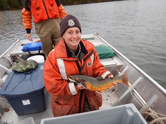
The Iron River National Fish Hatchery, WI maintains approximately 10,000 adult and juvenile captive lake trout and coaster brook trout brood stock. Infusion of wild genetics into these captive lines is paramount for successful brood stock management. This fall 2014, Carey Edwards and Brandon Keesler from Iron River and Henry Quinlan from the Ashland Fish and Wildlife Conservation Office, WI, made the trek to Isle Royale National Park in Lake Superior to collect gametes from coaster brook trout found in Tobin Harbor. Male gametes are collected in plastic bags and checked under a microscope for viability before collecting and fertilizing eggs from females. Once the eggs are fertilized with the milt, they are rinsed, disinfected, and sealed in containers with well water from the hatchery. The containers are packed in coolers for transport back to the mainland and continue on to a quarantine facility at Genoa Natrional Fish Hatchery, WI, to incubate, hatch and grow for 18 months before becoming brood stock at Iron River. USFWS photo.
http://www.fws.gov/midwest/ironriver/
http://www.fws.gov/midwest/ashland/
http://www.fws.gov/midwest/genoa/

Recommendation and review posted by Bethany Smith
Genetic Engineering Recast – Video
Genetic Engineering Recast
English 1001 recast project.
By: Brent Ufkes
View original post here:
Genetic Engineering Recast - Video
Recommendation and review posted by Bethany Smith
Caffeine counters cocaine's effects on women's estrus cycles
PUBLIC RELEASE DATE:
20-Nov-2014
Contact: Kathryn Ryan kryan@liebertpub.com 914-740-2100 Mary Ann Liebert, Inc./Genetic Engineering News @LiebertOnline
New Rochelle, NY, November 20, 2014-Women are more sensitive to the effects of cocaine and more susceptible to cocaine abuse than men. Cocaine's ability to disrupt a woman's estrus cycle may explain the sex differences in cocaine addiction, and new evidence that caffeine may be neuroprotective and able to block cocaine's direct effects on the estrus cycle reveals novel treatment possibilities, according to an article published in Journal of Caffeine Research: The International Multidisciplinary Journal of Caffeine Science, a peer-reviewed journal from Mary Ann Liebert, Inc., publishers. The article is available free on the Journal of Caffeine Research website at http://online.liebertpub.com/doi/full/10.1089/jcr.2014.0015 until December 20, 2014.
In the article "Cocaine Shifts the Estrus Cycle Out of Phase and Caffeine Restores It", Patricia Broderick, PhD and Lauren Malave, City College of New York, City University of New York Graduate Center, City University of New York, and NYU Langone Medical Center, New York, NY, show that cocaine shifts the estrus cycle, thereby changing a woman's estrogen levels. Caffeine can block these changes, suggesting that antagonists of the adenosine system may have a role in treating cocaine addiction.
"This is cutting-edge work that has never been shown before. It is critical knowledge relevant to women's reproductive health," says Patricia A. Broderick, PhD, Editor-in-Chief of Journal of Caffeine Research and Medical Professor in Physiology, Pharmacology & Neuroscience, The Sophie Davis School of Biomedical Education, The City College of New York, The City University of New York, and Adjunct Professor in Neurology, New York University Langone Medical Center and Comprehensive Epilepsy Center.
###
About the Journal
Journal of Caffeine Research: The International Multidisciplinary Journal of Caffeine Science is a quarterly journal published in print and online. The Journal covers the effects of caffeine on a wide range of diseases and conditions, including mood disorders, neurological disorders, cognitive performance, cardiovascular disease, and sports performance. Journal of Caffeine Research explores all aspects of caffeine science including the biochemistry of caffeine; its actions on the human body; benefits, dangers, and contraindications; and caffeine addiction and withdrawal, across all stages of the human life span from prenatal exposure to end-of-life. Tables of content and a sample issue may be viewed on the Journal of Caffeine Research website at http://www.liebertpub.com/jcr.
About the Publisher
More:
Caffeine counters cocaine's effects on women's estrus cycles
Recommendation and review posted by Bethany Smith
Panel-Based Genetic Diagnostic Testing for Inherited Eye Diseases Is Highly Accurate and More Sensitive Than Exome …
Contact Information
Available for logged-in reporters only
Newswise BOSTON (Nov. 20, 2014) Investigators at Massachusetts Eye and Ear and Harvard Medical School Department of Ophthalmology and colleagues reported the development and characterization of a comprehensive genetic test for inherited eye disorders in the online version of the Nature journal Genetics In Medicine today. The Genetic Eye Disease (GEDi) test includes all of the genes known to harbor mutations that cause inherited retinal degenerations, optic atrophy and early onset glaucoma. These disorders are important causes of vision loss, and genetic treatments such as gene therapy hold promise for preserving vision in affected individuals. The GEDi test is offered on a CLIA-certified basis through the Ocular Genomics Institute (OGI) at Mass. Eye and Ear.
The retina is the neural tissue in the back of the eye that initiates vision. It is responsible for receiving light signals and converting them into neurologic signals, which are then transmitted via the optic nerve to the brain so that we can see. Mutations that disrupt vision by damaging the retina and optic nerve have been identified in more than 200 genes. This genetic diversity made genetic diagnostic testing difficult until the recent development of high throughput genomic techniques. The GEDi test uses targeted capture and next generation sequencing techniques to sequence 226 genes known to cause inherited eye disorders. Future versions of the test will also include genes responsible for eye movement disorders (strabismus) and other inherited eye conditions.
Gene panel-based tests for inherited eye disorders have been previously reported, but none of these have been as thoroughly characterized with regard to their performance in a diagnostic setting as the GEDi test. Stringent tests of accuracy and reproducibility showed that the GEDi test is both highly accurate and reproducible. This type of validation testing is recommended by the American College of Medical Genetics and Genomics, but few other genetic tests have been characterized in as much detail as the GEDi test. The results reported show that the GEDi test is 98 percent accurate at detecting spelling variations or mutations in the genetic code of inherited eye disease genes, and is highly reproducible between test runs. In contrast, the technique whole exome sequencing in which the coding regions of all genes are sequenced, and which is being employed commonly in clinical settingswas 88 percent accurate at detecting genetic variants in the same genes.
The results we obtained for the GEDi test have broad implications and show that panel-based testing focused on the specific genes associated with genetic conditions offers important advantages over whole exome sequencing, said Janey Wiggs, M.D., Ph.D., director of the Genetic Diagnostic Testing Service of the OGI, and the Paul Austin Chandler Associate Professor of Ophthalmology, Harvard Medical School.
Investigators in the OGI and other centers around the United States and the world are optimistic that treatments targeting the underlying genetic cause of inherited eye disorders can be applied broadly to preserve vision. One especially promising approach is gene therapy, in which a correct copy of the misspelled or mutant gene responsible for disease is added to the affected cells in the retina. Reports of early results from clinical trials of gene therapies for two inherited retinal degenerative disorders have shown that this treatment can be performed safely, and that subjects treated in these trials experienced significant improvements in or preservation of vision. Clinical trials of gene therapies for three additional genetic forms of inherited retinal degeneration are currently in progress, and more are on the way. Given the potential of gene and genetic therapies, improved genetic diagnostic testing for patients with genetic eye disorders such as that offered with the GEDi test is especially important.
About Massachusetts Eye and Ear Mass. Eye and Ear clinicians and scientists are driven by a mission to find cures for blindness, deafness and diseases of the head and neck. After uniting with Schepens Eye Research Institute in 2011, Mass. Eye and Ear in Boston became the world's largest vision and hearing research center, offering hope and healing to patients everywhere through discovery and innovation. Mass. Eye and Ear is a Harvard Medical School teaching hospital and trains future medical leaders in ophthalmology and otolaryngology, through residency as well as clinical and research fellowships. Internationally acclaimed since its founding in 1824, Mass. Eye and Ear employs full-time, board-certified physicians who offer high-quality and affordable specialty care that ranges from the routine to the very complex. U.S. News & World Reports Best Hospitals Survey has consistently ranked the Mass. Eye and Ear Departments of Otolaryngology and Ophthalmology as among the top hospitals in the nation. Mass. Eye and Ear is home to the Ocular Genomics Institute which aims to translate the promise of precision medicine into clinical care for ophthalmic disorders. For more information about life-changing care and research, or to learn how you can help, please visit MassEyeAndEar.org.
Reference: Consugar MB*, Navarro-Gomez D*, Place EM*, Bujakowska KM, Sousa ME, Fonseca-Kelly ZD, Taub DG, Janessian M, Wang DY, Au ED, Sims KB, Sweetser DA, Fulton AB, Liu Q, Wiggs JL,Gai X, Pierce EA. Panel-based Genetic Diagnostic Testing for Inherited Eye Diseases is Highly Accurate and Reproducible and More Sensitive for Variant Detection Than Exome Sequencing. Genetics In Medicine, In Press. (*Co-first authors).
Grant support: This work was supported by grants from the National Institutes of Health (EY012910, and P30EY014104), the March of Dimes and the Foundation Fighting Blindness.
See the original post:
Panel-Based Genetic Diagnostic Testing for Inherited Eye Diseases Is Highly Accurate and More Sensitive Than Exome ...
Recommendation and review posted by Bethany Smith
Veterinary Pharmacology Research Foundation Continues Support for American College of Veterinary Internal Medicine
Contact Information
Available for logged-in reporters only
Newswise Denver, Colorado -- The American College of Veterinary Internal Medicine Foundation (ACVIMF), in partnership with the Veterinary Pharmacology Research Foundation (VPRF), announced today that they have funded a new study for animal health with a pharmacologic focus. The study will be conducted by Dr. Lauren Trepanier of the University of Wisconsin-Madison for the Genetic risk for cyclophosphamide toxicity in dogs.
The strategic partnership between VPRF and ACVIMF began in 2009 in an effort to solicit, review and administer research grants with a pharmacologic focus. The Veterinary Pharmacology Research Foundation (VPRF) was formed by the governing bodies of American Academy of Veterinary Pharmacology and Therapeutics (AAVPT) and the American College of Veterinary Clinical Pharmacology (ACVCP) in June of 2006. These organizations recognized that the lack of funding for basic pharmacology research was limiting both growth and innovation in the development of new veterinary therapeutics and the number of trained researchers in the field. As such, they sought to invest their collective resources in the growth of veterinary pharmacology through research grants and training programs. Jane Owens, President, VPRF.
The first call for proposals went out in November 2009. Since then, the partnership has awarded over $88,250 to six ACVIM Board-certified researchers along with their research team members. The first VPRF grant was awarded in June 2010 to Drs. Kenneth Simpson, Melanie Craven and Belgin Dogan from Cornell University for the development of a novel amikacin delivery method for treatment of E. coli associated with Granulomatous Colitis of Boxer dogs.
In 2011, the second grant was awarded to Drs. Butch KuKanich and Kate KuKanich from Kansas State University for a study to determine the effect of CYP inhibition on tramadol disposition and pharmacological effects in dogs. In 2012, Drs. Chen Gilor and Christopher Adin of the Veterinary Clinical Sciences Department at the Ohio State University received funding for evaluating Exenatide extended release in cats. Grant monies were also awarded to Drs. Jennifer Myers, Janice Bright, Christopher Orton, Daniel Gustafson and Christine Swardson Olver from the College of Veterinary Medicine & Biomedical Sciences, Colorado State University for evaluation of the pharmacokinetics and pharmacodynamics of Apibaxin in cats. The 2013 VPRF funds were awarded to Dr. Dawn Boothe and resident Jacqueline Gimmler of Auburn University for establishing terbinafine doses for treatment of canine Malassezia infection.
The grants encourage investigators to submit proposals that focus on research to evaluate the safety, effectiveness and duration of effect of therapies for veterinary species, explore new drug therapies for animals, develop and validate methods of evaluating effects of drugs in animal diseases or conditions, or ensure that a safe food supply is not compromised by drug therapy. As this grant is a partnership between veterinary internists and VPRF, collaborations between pharmacologists and Diplomates of ACVIM were strongly encouraged.
About Veterinary Pharmacology Research Foundation The Veterinary Pharmacology Research Foundation provides grant funding to support research into new and currently approved medications for combating diseases of companion and food animals, projects that ensure the safety of food products from treated livestock, and training programs for veterinary pharmacologists. These areas have been unmet needs in veterinary medicine for over 30 years. To donate to the Foundation, please access the following: http://aavpt.affiniscape.com/associations/12658/files/VPRFDonationForm3.pdf
About the ACVIM Foundation The American College of Veterinary Internal Medicine (ACVIM) Foundation is a non-profit 501(c)(3) organization dedicated to pioneering the healthcare of animals through the work of specialists in the American College of Veterinary Internal Medicine (ACVIM): small and large animal internists, cardiologists, neurologists, and oncologists. By supporting the work of these groundbreaking scientists, by raising awareness of specialty medicine, and by mobilizing the animal-loving public, we aim to revolutionize current treatments and spark the discovery of future cures. The ACVIM Foundation recognizes the need for advanced care, research dollars, awareness, and the need to support the Resident-in-training and the future scientist. Learn more at http://www.ACVIMFoundation.org.
# # #
Continued here:
Veterinary Pharmacology Research Foundation Continues Support for American College of Veterinary Internal Medicine
Recommendation and review posted by Bethany Smith
Confusion Of Faces and Genetics – Video
Confusion Of Faces and Genetics
A child #39;s nationality is based off the father because the man carries the seed.
By: GMSNebraskaWatchmen5
See the rest here:
Confusion Of Faces and Genetics - Video
Recommendation and review posted by Bethany Smith
Chiara Bonini: "Bases of Gene Therapy in leukemias" – Video
Chiara Bonini: "Bases of Gene Therapy in leukemias"
Chiara Bonini, dept. of Experimental Hematology at Fondazione San Raffaele del Monte Tabor - Milano, talks about "Bases of Gene Therapy in leukemias". http:/...
By: European Society for Gene and Cell Therapy
Go here to see the original:
Chiara Bonini: "Bases of Gene Therapy in leukemias" - Video
Recommendation and review posted by Bethany Smith